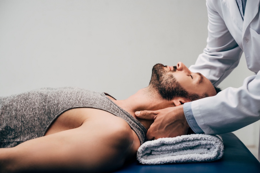
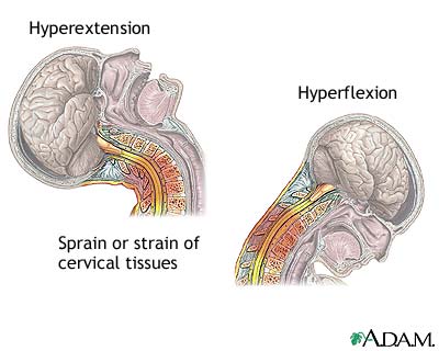
As,we know that with aging process cervical spine muscles degenerates so,it affects person daily activities which leads to severe cervical neck pain and stretching of neck muscles which looses its flexibility and strength.
Here are some tests that are prescribed by Radiologists to, investigate radiological abnormalities which occur due to cervical spine degeneration:
1.X-Ray 2.MRI 3.CT Scan 4.Blood Tests 5.Bone Density Assessment
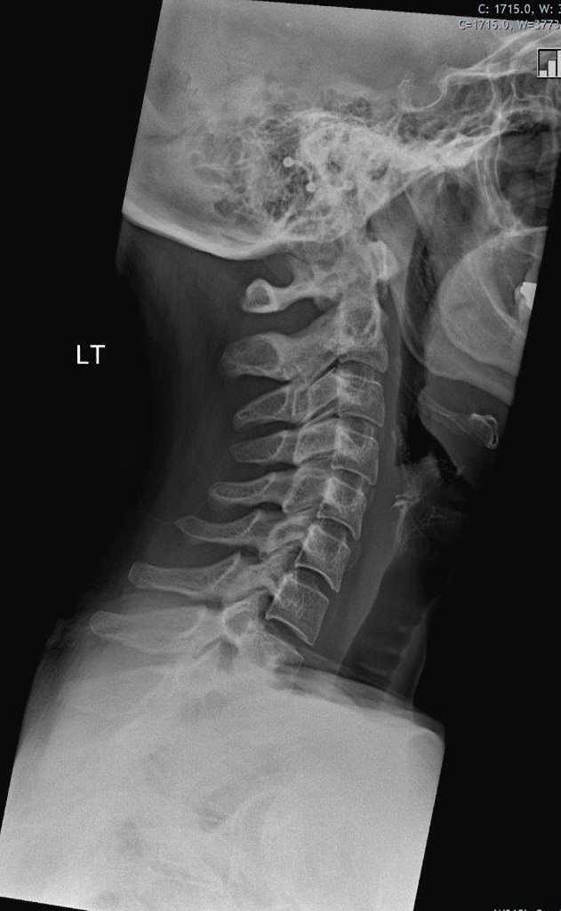
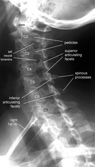
1.X-Ray:
It is easiest mean to image the spine.X-ray reveals alignment and degenerative changes of the bones.The spaces for the discs are seen as well, but no pictures are seen of the spinal cord,nerves or actual disc material. Unsuspected bony pathology, such as fractures,dislocations and cancer metastases, are quickly identified with X-ray.
X-RAY
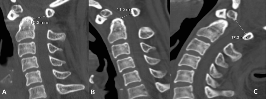
2.CT-Scan:
It is useful for cross-sectional imaging of the spine and increased image detail of the spinal cord,nerves and discs are investigated.
CT-SCAN
3.MRI Imaging:
MRI is currently the best means of all of the important structures of the cervical spine.With a good MRI,considerable detail is available of the bones,discs,spinal cord, ligaments and the nerves.
MRI
4.Blood Work:
It is done,if suspicion of spinal cord disease is present.Also,rheumatoid arthritis may be detected with blood work.
5.Bone Density Assessment:
It assists in diagnosis of a loss of calcium(Ca+) as seen in osteopenia and osteoporosis,conditions that weaken the bone structure everywhere.
TESTS(ASSESSMENT FOR CERVICAL NECK PAIN)
Everyone is different, and your specific neck condition may require unique exercises that are different as everyone requires different assessments,if radiological findings are abnormal.
As,daily living,poor posture,and injury often result in tight neck muscles.If you have neck pain or tightness in your neck muscles,physical therapist can help prescribe the best exercises to help improve your ability to move your neck fully and with little or no pain.Stretching your neck can have a positive impact on your pain, posture,and overall mobility.
There are four gentle neck stretching exercises that physical therapist prescribe to decrease muscle tightness.
Usually,stretching exercises can be performed while lying on your back (to provided assisted support) or in the sitting or standing position.Physical Therapist prescribe their patients to do exercises at home to get better results.
Be sure to check in with physical therapist before starting these,or any other,neck exercises.
EXERCISES:
Following exercises are mentioned below:
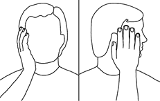
1.Flexion Stretch:Chin to your chest.
Starting Position:Begin each exercise with your neck in midline position. Your head should be centered and not tilted forward, back or to the side.You can do this exercise while either
FLEXION STRETCH
Position:Lying flat on your back or sitting on seat.
Practical:Gently bend your head forward while bringing your chin toward your chest. Stop when a stretch is felt in the back of your neck. Hold position for 20 seconds.
Repetition:Repeat this four more times for a total of five repetitions.
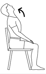
2.Cervical Extension/Extension Stretch:Eyes Upto the Sky.
Starting Position:Performer is looking up and it can help relieve tension in your neck.Extension of your cervical spine may also be useful to help relieve pain from bulging discs in your neck.
CERVICAL EXTENSION STRETCH
Position:Lying flat on your back or sitting up.
Practical:Gently bend your head backward so that your eyes are looking up. Stop when a stretch is felt in the front of your neck. Hold position for 20 seconds.
Repetition:Repeat this four more times.
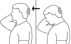
3.Rotation:Side to Side.
Starting Position:Begin each exercise with your neck in midline position.
ROTATION STRETCH
Position:Lying flat on your back or in sitting.
Practical:Gently turn your head to the left, looking over your left shoulder. Stop when a stretch is felt in the right side of your neck. Hold position for 20 seconds.
Repetition:Repeat above stretch four times.
*This stretch can be perform in opposite direction i.e:if your are turning right to left or left to right.
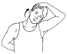
4.Lateral Flexion:Ear to Shoulder.
Starting Position:Head should be centered and not tilted forward, back, or to the side.
LATERAL FLEXION STRETCH
Position:Lying flat on your back or sitting up.
Practical:Bend your neck in attempts to touch your left ear to your shoulder. Stop when a stretch is felt in the right side of your neck. Hold position for 20 seconds.
Repetition:Repeat above stretch four more times.
*This stretch can be perform in opposite direction i.e:if your are turning right to left or left to right.
By performing,these exercises regularly or twice or thrice a week, this can help patients to get better and desirable results.
Although,therapeutic techniques are mostly used such as rest, massage,ice and heat which are certainly the first step in treating the acute onset of cervical spine pain,particularly if muscular in origin. Sometimes several weeks are necessary for resolution.If the symptoms seem to be gradually improving,no medical intervention may be necessary.Avoiding poor neck posture and positions is essential.Sitting up straight and keeping the head centered over the chest while sitting improves posture.
TREATMENT:
So,the helpful modifications include:
(1)Avoid working with the arms up overhead or the neck in a persistently flexed position.
(2)Avoid frequent twisting or turning.
(3)Avoid carrying a computer or heavy bag over one shoulder or cradling the phone to one ear.
(4)Try to sleep with fewer pillows and avoid sleeping on one’s stomach.Massage may loosen up the muscles. Ice may calm the inflammation at the onset of difficulties and heat may later relax the muscles.
CERVICAL NECK STRETCH
SEVERITY OF NECK PAIN:
Neck Pain may be chronic or recent in onset.It may be confined to the neck or radiate to the arms.
Such,as mild or severe and dull or sharp and better or worse with certain physical activities.
a)MILD STAGE:
Headache is usually daily,in back of the skull and radiates forwards over the temple,mild and relieved with minor pain medications.
b)CHRONIC STAGE:
It can be quite severe and mistaken for “migraine”.
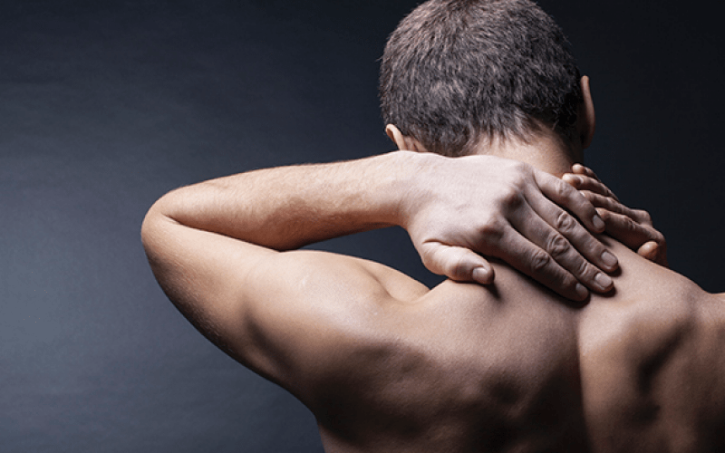
CERVICAL NECK PAIN
FACTORS AFFECTING NECK PAIN:
Factors affecting cervical neck pain are mentioned below:
1.Numbness into the arm.
2.Fatigue of certain motions may be specifically recognized and reported as weakness.
3.Bowel,bladder,gait and balance difficulties gives red flags to spinal cord injury.
4.Stress management may not be linked to psychological tension as it affects neck region and work stress with long hours at the computer or desk.
5.Poor sleep often results in stress as the neck muscles don’t get relax well when sleep is poor.
6.Anti-inflammatory agents,analgesics and muscle relaxants work best for most spine discomfort.Such as, ibuprofen,naproxen and aspirin.
In the last,this approach is for temporary period until the anatomy and mechanics of the spine dysfunction are identified and corrected.
Neck pain which is also called as Cervical Neck pain. In this, physiotherapy can have a massive impact on the global burden of neck pain.Through the application of good examination skills, effective clinical reasoning and appropriate selection of interventions the impact of neck pain for any individual can be significantly reduced. In addition the high tendency for chronicity in neck pain can be lowered with effective management.
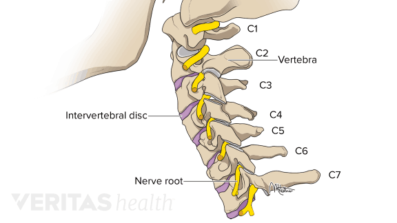
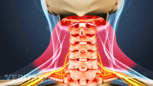
Cervical Vertebrae(C1-C7)
INTRODUCTION:
The cervical spine is the most superior portion of the vertebral column, lying between the cranium and the thoracic vertebrae.
It consists of seven distinct vertebrae, two of which are given unique names:
1.ATLAS:
The first cervical vertebrae (C1) is known as the atlas.
2.AXIS:
The second cervical vertebrae (C2) is known as the axis.
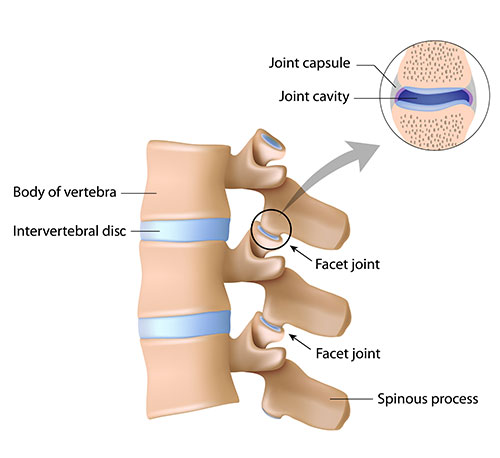
ATTACHMENT: Attached to the back of each vertebral body is an arch of bone that forms a continuous hollow longitudinal space, which runs the whole length of the back. This space, called the spinal canal, is the area through which the spinal cord and nerve bundles pass. The spinal cord is bathed in cerebrospinal fluid (CSF) and surrounded by three protective layers called the meninges (dura, arachnoid, and pia mater).
VERTEBRAL LEVEL:
At each vertebral level, a pair of spinal nerves exit through small openings called foraminae (one to the left and one to the right). These nerves serve the muscles, skin and tissues of the body and thus provide sensation and movement to all parts of the body. The delicate spinal cord and nerves are further supported by strong muscles and ligaments that are attached to the vertebrae.
CERVICAL NECK PAIN
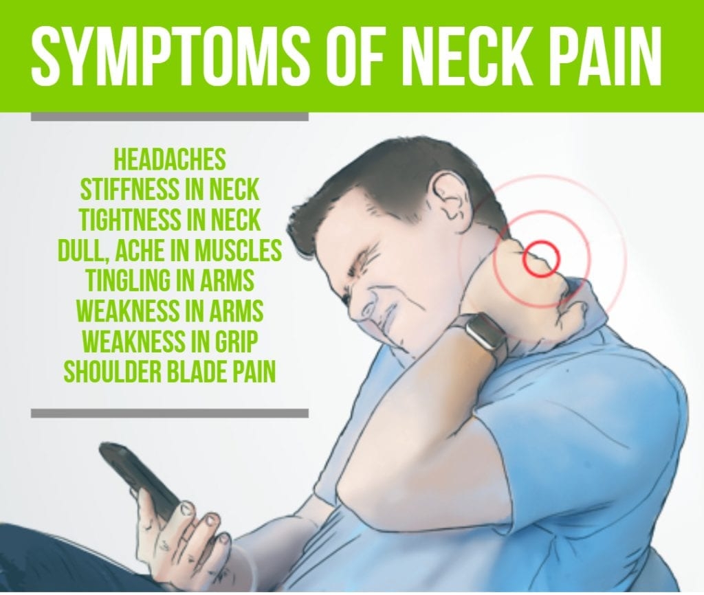
SYMPTOMS OF NECK PAIN:
Neck pain of soft tissue origin tends to produce symptoms localized to the muscular tissues running up and down both sides of the cervical spine. The discomfort can expand out towards the shoulders, but rarely radiates down the arms.
SYMPTOMS:
They are commonly worse with the head and neck in a fixed, or static, position and better with movement, ice or heat and massage. This tends to be a persistent type of pain, often precipitated by injury or overuse. The cause can be injuries to the muscles and ligaments or to the underlying bones and discs. Once injury occurs, there tends to be a tightening of the muscles, perhaps as a reflex designed to hold the head and neck stable, but this also creates enhanced muscle tone and discomfort from the chronic muscle spasm.
SYMPTOMS
Motor Function:
Almost all of the muscles in both the arms and legs are tested.Maximum power that each muscle can generate and the loss of muscle bulk (atrophy) are assessed.
Sensory Function:
It is tested with either a pin-prick or light-touch method, looking for areas of numbness, tingling or burning.
Reflex Activity:
The arms and legs are tested with the rubber hammer to provide insight to nerve, spinal cord and muscle function.
Gait Assessment And Coordination:
It is checked through balance and pattern of muscle power. Coordination of both arms and legs is reviewed for both dexterity and balance.
Range of Motion:
Mostly all of the spine, both passively and actively, is performed while assessing the musculature and identifying whether any nerve, spinal cord or pain difficulties emerge.
To preserve flexibility and strength of Achilles Tendon,a treatment plan which is prescribed by physiotherapist and patient has to follow the plan at home to maintain their health and fitness to keep them active and healthy.
Here are some of the stretches that will maintain flexibility and restoration of rupture tendon:
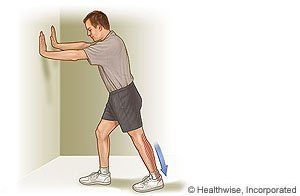
1.Floor Stretch:
Method:
1.Stand about 0.5 metres from a wall, and place your hands on the wall at about shoulder height or you can stand behind a chair, placing your hands on the back of it for balance. 2.Step back with the leg you want to stretch. Keep the leg straight, and press your heel into the floor with your toe turned slightly in. 3.Lean forward, and bend your other leg slightly. Feel the stretch in the Achilles tendon of your back leg.
*Hold for at least 15 to 30 seconds. Repetition:2 to 4 times a session, up to 5 sessions a day.
FLOOR STRETCH
2.Stair Stretch:
Method:
1.Stand with the balls of both feet on the edge of a step or curb (or a medium-sized phone book).
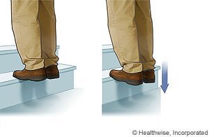
2.With at least one hand, hold onto something solid for balance, such as a banister or handrail. 3.Keeping your affected leg straight, slowly let that heel hang down off of the step or curb until you feel a stretch in the back of your calf and/or Achilles area. Some of your weight should still be on the other leg. *Hold this position for at least 15 to 30 seconds. Repetition:2 to 4 times a session, up to 5 times a day or whenever your Achilles tendon starts to feel tight.
STAIR STRETCH
3.Strength Exercise:
Method:
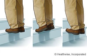
1.This exercise will get you started on building strength after an Achilles tendon injury.Your physiotherapist can help you move on to more challenging exercises as you heal and get stronger. 2.Stand on a step with your heel off the edge of the step. Hold on to a handrail or wall for balance. 3.Push up on your toes, then slowly count to 10 as you lower yourself back down until your heel is below the step. Repetition: Repeat exercise 8 to 12 times, half with the knee straight and half with the knee bent.
STRENGTH EXERCISE
4.Toe Stretch:
Method:
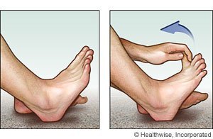
1.Sit in a chair, and extend your affected leg so that your heel is on the floor. 2.With your hand, reach down and pull your big toe up and back. Pull toward your ankle and away from the floor. *Hold the position for at least 15 to 30 seconds. Repetition: 2 to 4 times a session, several times a day.
TOE STRETCH
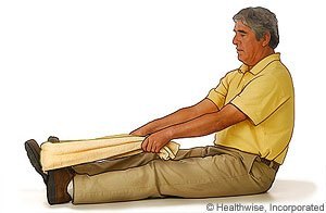
5.Calf Plantar Fascia Stretch:
Method:
1.Sit with your legs extended and knees straight. 2.Place a towel around your foot just under the toes. 3.Hold each end of the towel in each hand, with your hands above your knees. 4.Pull back with the towel so that your foot stretches toward you. *Hold the position for at least 15 to 30 seconds. Repetition: 2 to 4 times a session, up to 5 sessions a day.