As,we know that with aging process cervical spine muscles degenerates so,it affects person daily activities which leads to severe cervical neck pain and stretching of neck muscles which looses its flexibility and strength.
Here are some tests that are prescribed by Radiologists to, investigate radiological abnormalities which occur due to cervical spine degeneration:
1.X-Ray 2.MRI 3.CT Scan 4.Blood Tests 5.Bone Density Assessment
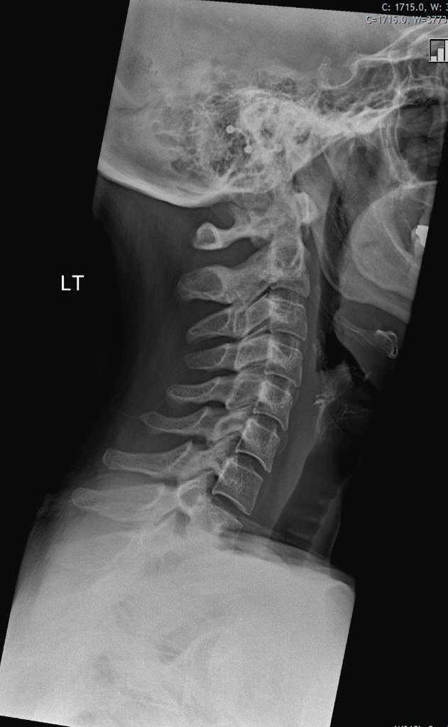
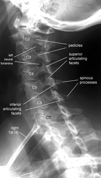
1.X-Ray:
It is easiest mean to image the spine.X-ray reveals alignment and degenerative changes of the bones.The spaces for the discs are seen as well, but no pictures are seen of the spinal cord,nerves or actual disc material. Unsuspected bony pathology, such as fractures,dislocations and cancer metastases, are quickly identified with X-ray.
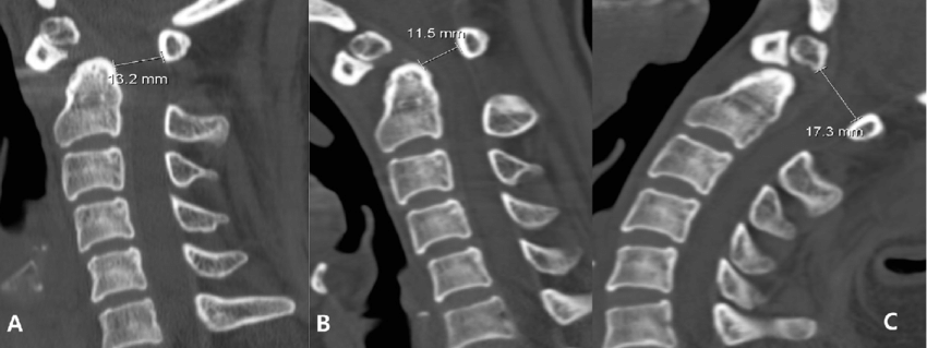
2.CT-Scan:
It is useful for cross-sectional imaging of the spine and increased image detail of the spinal cord,nerves and discs are investigated.
3.MRI Imaging:
MRI is currently the best means of all of the important structures of the cervical spine.With a good MRI,considerable detail is available of the bones,discs,spinal cord, ligaments and the nerves.
4.Blood Work:
It is done,if suspicion of spinal cord disease is present.Also,rheumatoid arthritis may be detected with blood work.
5.Bone Density Assessment:
It assists in diagnosis of a loss of calcium(Ca+) as seen in osteopenia and osteoporosis,conditions that weaken the bone structure everywhere.
Everyone is different, and your specific neck condition may require unique exercises that are different as everyone requires different assessments,if radiological findings are abnormal.
